The HyProCure Advantage
What is The HyProCure Advantage
HyProCure is the proven leader in the extra-osseous talotarsal stabilization arena. There is no other treatment option that comes even close to the results of HyProCure. Other sinus tarsi implants may make unsubstantiated claims that they are the same as HyProCure, but they don’t have the scientific evidence to prove it. HyProCure is back by an extensive pedigree of published studies that have established it as the best sinus tarsi spacer.
HyProCure’s success is based on its anatomic design and biomechanic function.
Need more proofs?
Check out the list of publications of/on HyProCure I and II
The top 5 options for the treatment of ankle bone instability are:
- Observation – the “do-nothing” approach
- Arch supports/foot orthotics
- Traditional rearfoot reconstructive surgery – the nuclear options
- Type I arthroereisis devices (non-HyProCure designed implants)
- HyProCure extra-osseous talotarsal stabilization device
Observation – Guaranteed to eventually lead to tissue disease
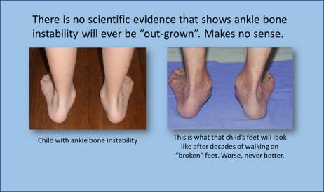
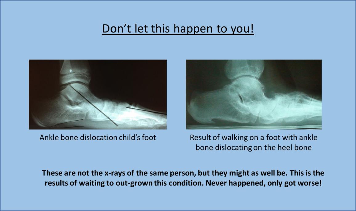
There are many physicians who downplay or simply ignore ankle bone instability as a normal variant. They will tell their patients not to worry about those misaligned feet. Meanwhile, every step taken is a one step closer to painful conditions within the feet, knees, hips, or back. Not everyone with ankle bone instability will have a painful foot, but painful symptoms will eventually show up sooner than later. It is just a matter of time. Initially, it may seem like there is no rush to have the ankle bone internally stabilized, and yes this is not a life-threatening condition, but don’t wait too long. The secondary tissues could have been saved if caught early, otherwise there is a point of no return were that tissue will become permanently diseased.
Initially, there will be functional symptoms. The more active you are the more you suffer. Then those painful areas to the feet, knees, hips or back will need someone kind of treatment, but those treatment will eventually fail. The leading long-term problem with surgery to your feet, knees, hips and back is that it’s only a matter of time until more surgery is needed. That’s because the underlying cause is still present. Yes, that’s right, that ankle bone instability is present and is still cause excessive forces to the body.
We are not trying to use scare tactics, but we need to make sure that you understand the fact that ankle bone instability only gets worse, never better.
Arch supports/Foot Orthotics
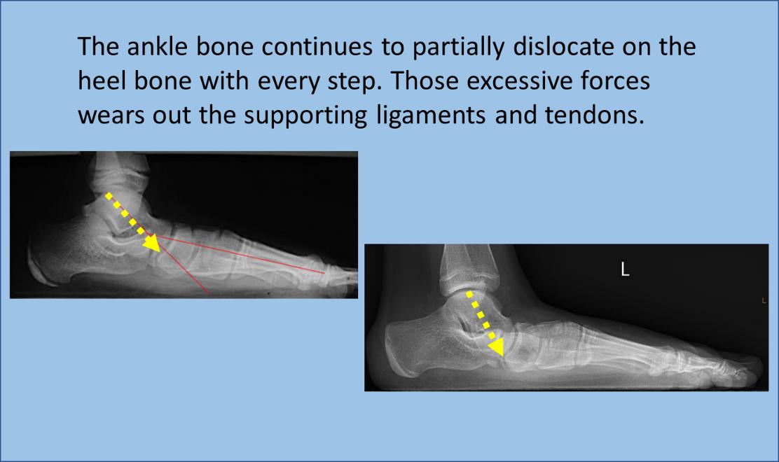
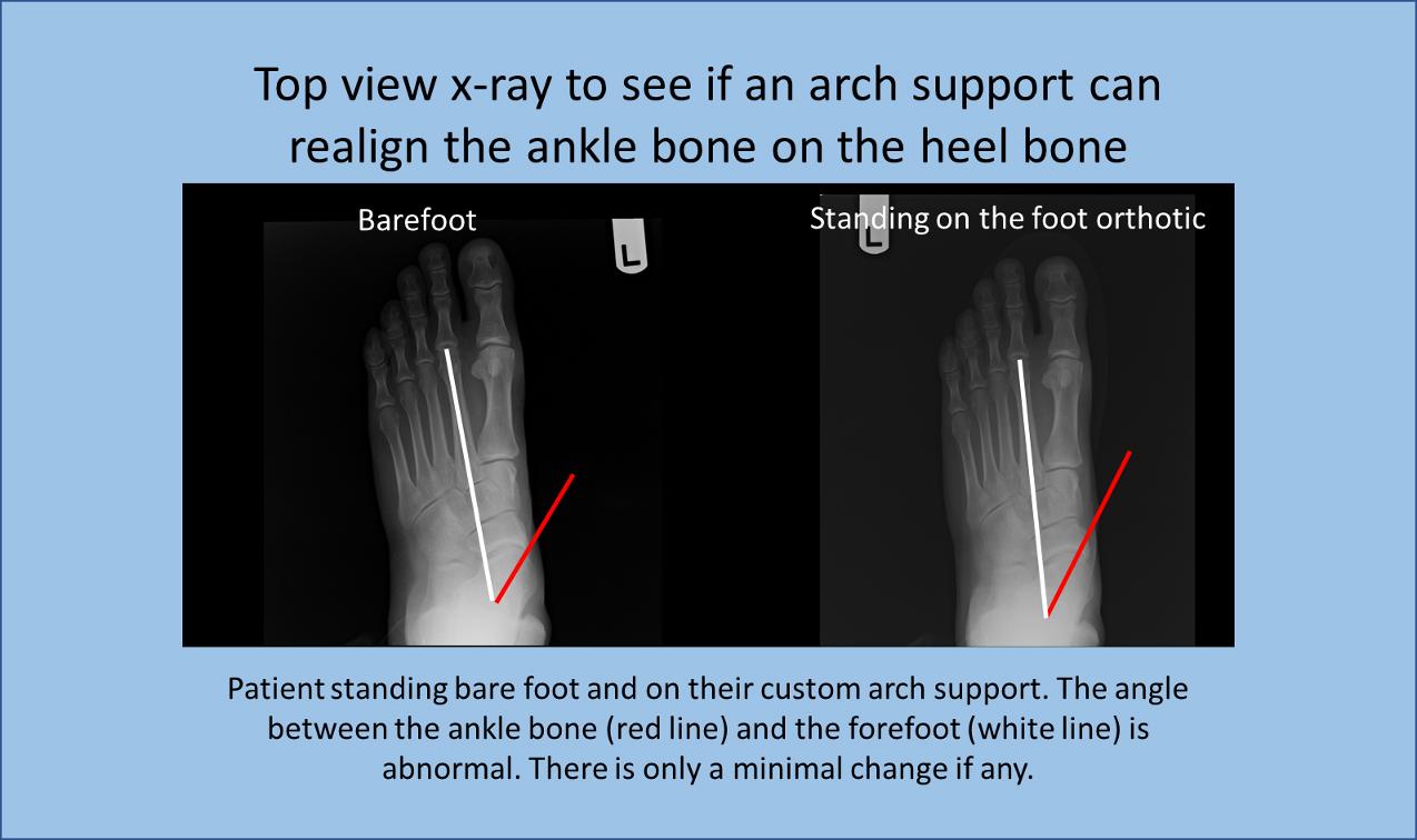
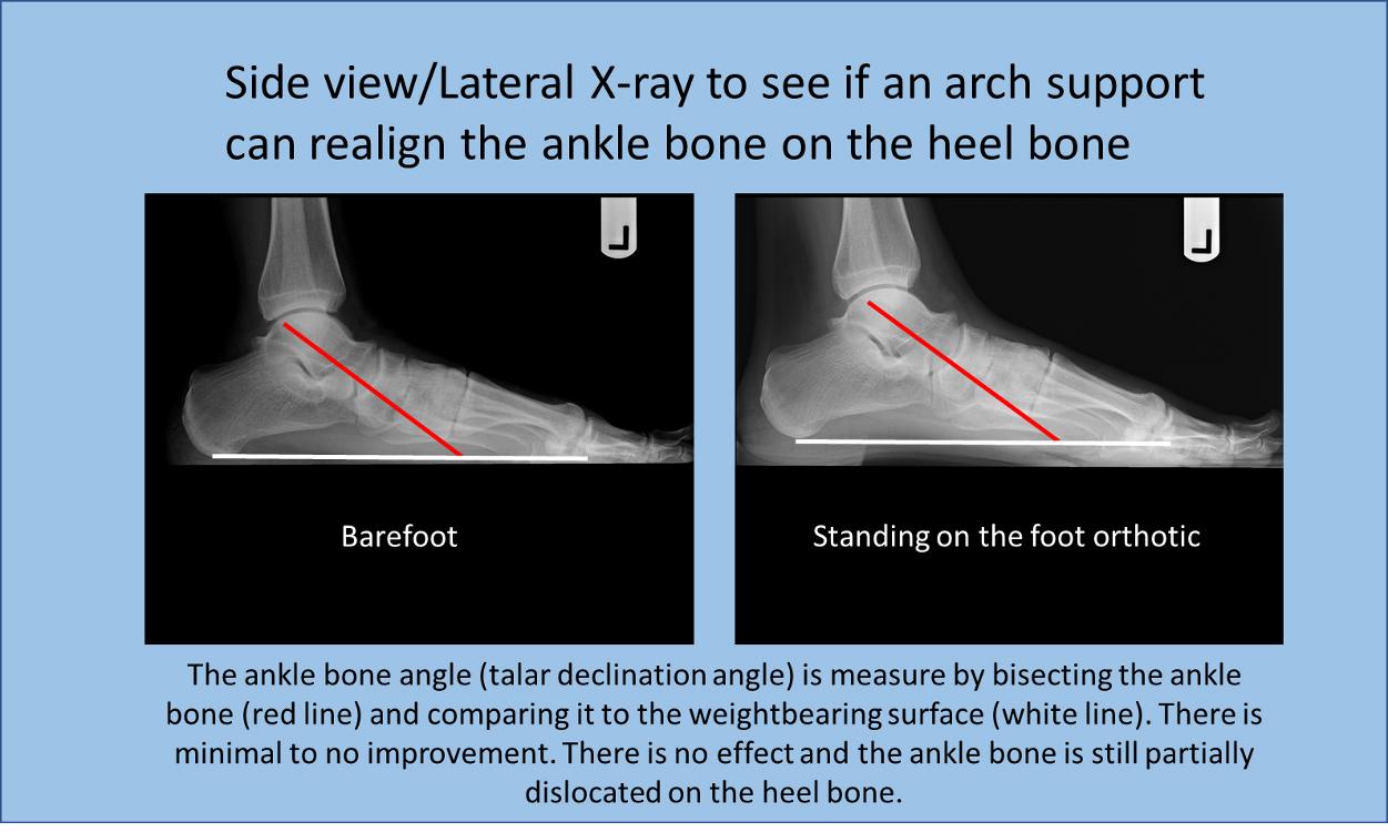
It is true that just about everyone could benefit from an insert in their shoe. Shoes are not friendly to our feet and the “arch” supports are usually just an optical illusion. There are many people whose feet feel better as a result of wearing arch supports. With that said, there is no possible way for an arch support, even the ones costing over $1000 of your hard-earned money, could ever stabilize and realign the ankle bone. That’s because this is an internal deformity and a non-surgical method cannot have a positive effect. Even if you had a walking cast placed on your foot and ankle, internally the ankle bone would still be partially dislocating on the heel bone.
Here is a simple test to see if your arch supports are realigning your feet, have your doctor take x-rays of your feet without the arch support and take another set standing on the arch support. There are 2 main angles the doctor should measure. The ankle bone declination angle (talar declination angle) on the side view, and the ankle-forefoot angle (talar 2nd metatarsal angle). You will find that there will be minimal changes in the angles if any.
Many people are told that their arch supports are fixing their misaligned, over-pronating feet but the reality is that they are not. Some may say what’s the harm in selling these devices to people with ankle bone instability. Well, it is great harm and should be considered below the standard of care. That’s because the disease entity is still present and while standing and with every step taken, excessive abnormal forces are continuing to destroy the bones, joints, ligaments, tendons, and nerves within the feet, knees, hips and back.
Simply put, the use of arch supports in the treatment of ankle bone instability should be considered a subtherapeutic form of treatment and therefore below the orthopedic standard of care.
Traditional Bone Reconstructive Surgery
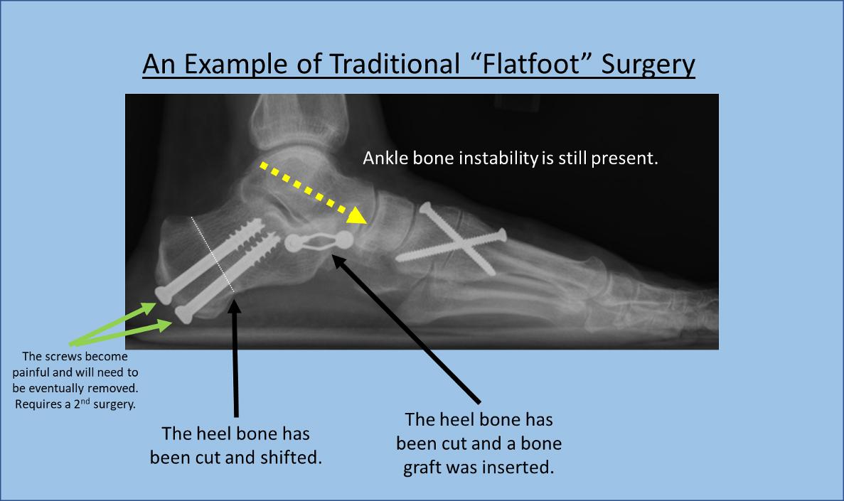
There are a variety of orthopedic surgeries that involve the cutting and shifting of bones to cutting out of a joint and fusing the two bones together in an attempt to stabilize the hindfoot. Unfortunately, for individuals who waited too long to have a more conservative procedure, like the insertion of HyProCure, this is now their only option.
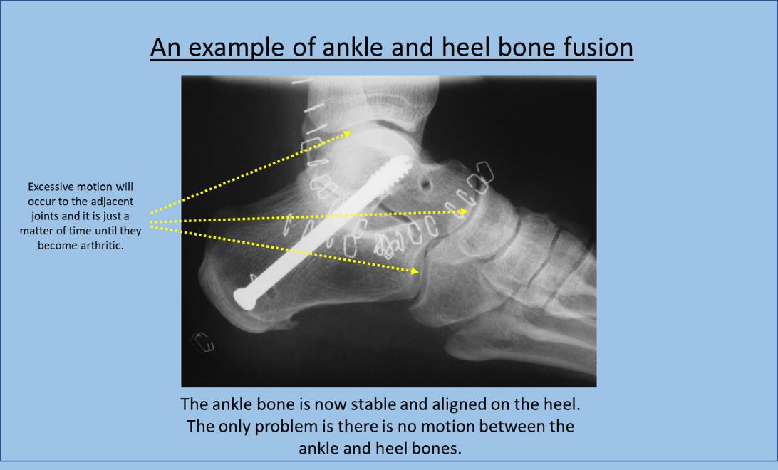
The issue with this surgical option is that once the bones are cut and shifted or fused, there is no going back. This is an irreversible treatment option. Also, these procedures are not mechanically friendly, they are the opposite – unfriendly. That’s because when a joint is removed, the motion that was supposed to occur in the joint has to now occur in the adjacent joint.
There is one procedure called a heel lengthening procedure (lateral column, or Evans procedure) where a bone graft is inserted to make the outer heel longer. A study showed that withing 6 months there were arthritic changes in the joint next the heel and outer foot bone (doi: 10.1177/1071100712464211).
Another procedure cuts and shifts the back the heel bone to correct an abnormal angulation. The problem with this procedure is that it does nothing to address the ankle bone instability issue. The head of the screw(s) that is inserted to hold the bones in place while they heal usually become painful and require a 2nd surgery.
There is another very aggressive surgery where the largest of the three joint surfaces between the ankle and heel bones is removed. This procedure is called a subtalar joint arthrodesis. The motion between the ankle and heel bones is completely eliminated. That means excessive motion will now occur within the ankle joint above and the bones in front of the ankle and heel bones. There has to be a better option!
Arthroereisis with an Intra-osseous Talotarsal Joint Stabilization Device (IOTTS)
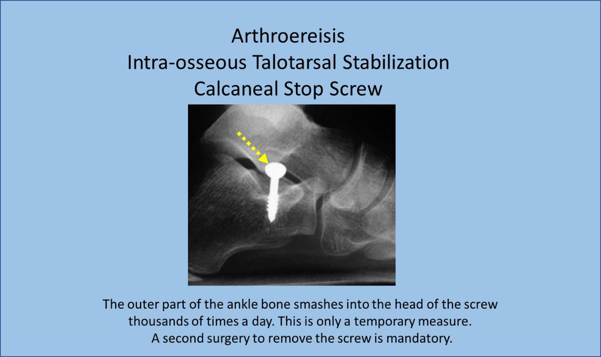
This procedure was developed as an option to stabilize the ankle bone without having to cut into bones or eliminate the joint. This procedure is called arthroereisis, (pronounced: r-throw-ear-e-sis). It literally means a joint blocking procedure. An implant, usually an inexpensive bone screw is just partially inserted into the heel bone. The top of the screw acts as a doorstop to prevent the ankle bone from sliding forward.
At first glance this seems like an economical, effective option but let’s explore this option a little more to discover it makes absolutely no sense.
First of all, this procedure is only recommended for those aged 14 to 18. That means you have to walk on deformed feet for up to several years until you reach 14 or if you are older than 18, sorry you need to try something else.
Think about how this works. When the foot is in the air during the walking cycle, the ankle bone is aligned on the heel bone. It’s when the heel touches the ground that the outer ankle bone slides forward and smashes into the head of the screw. This trauma occurs thousands of times a day and tens of thousands of time a week, hundreds of the thousands of times a month. That is why the screw must be removed.
Authors have claimed that the ankle bone continues to be stabilize after the removal of the screw, but there are no published radiographic studies to prove that fact. It really doesn’t make any sense. What was stabilizing the ankle bone has now been removed.
Advocates of this procedure say that it is better than other technique because the price of the screw is less expensive than other types of sinus tarsi spacers/stents. This seems to make sense because a bone screw is rather inexpensive. The big issue is that they don’t seem to factor in the added cost of having to remove the screw. The cost of having to remove the screw has to be more than the cost of a sinus tarsi spacer.
Let’s think about why this option makes little sense. A surgery was performed, the ankle bone was stabilized, but then screw had a be removed within 18 months. This option guarantees 2 surgical procedures, 2 trips to the operating room, and 2 times your child has to be put under general anesthesia. This means more expense and time and there is no evidence the ankle bone will remain stabilized. That really doesn’t make any sense, does it?
Arthroereisis with Extra-osseous Talotarsal Stabilization (EOTTS) Implant
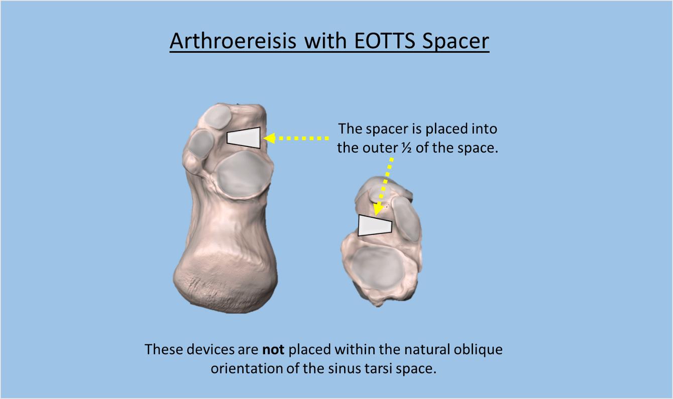
This option is an improvement over the IOTTS partial insertion of a screw into the heel bone. EOTTS involves the insertion of a spacer into the outer portion of the sinus tarsi space. The function is the same as the IOTTS without the need to drill into the heel bone. The device is held in place by soft tissues growing into and around the spacer.
There are many of these arthroereisis EOTTS spacers approved for use. they can remain in place longer because they are not drilled into bone. These devices are used in both children and adults, they do not have the age restrictions like the IOTTS devices.
The biggest issue with this arthroereisis device is that there are patients who eventually develop a soreness/pain from spacer and require removal. Some studies show removal rates greater than 30% (doi: 10.1053/j.jfas.2011.10.019, DOI: 10.1177/107110070602700103, DOI: 10.3113/FAI.2011.1127)
Non-Arthroereisis Extra-osseous Talotarsal Stabilization Device HyProCure
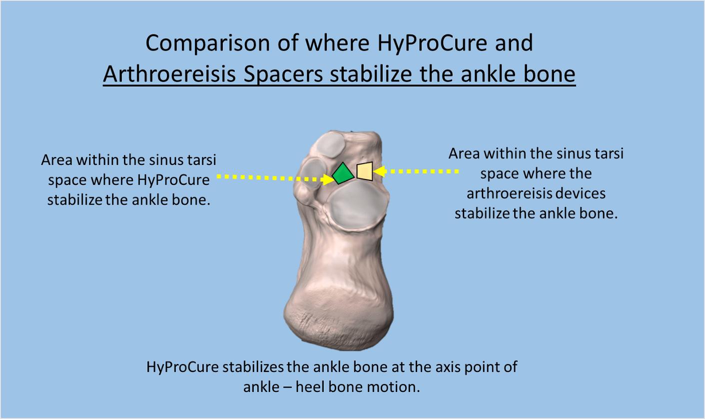
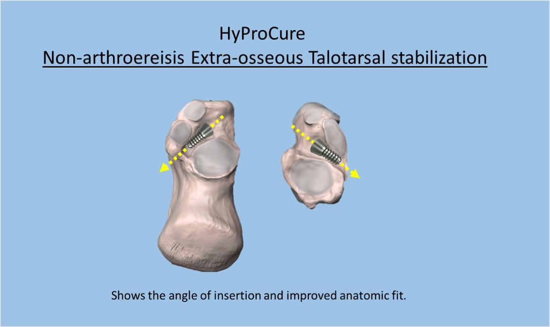
HyProCure was created to overcome the limitations of the arthroereisis types of spacers. A critical analysis by Dr. Graham showed the ideal shape of the spacer should closely mimic the space and alignment/orientation of the sinus tarsi. Also, rather that the ankle bone hitting up against the device as a bone blocking procedure, HyProCure should be designed so that the ankle bone would glide over HyProCure. The anatomic shape of HyProCure was independently analyzed that proved HyProCure had the best anatomic fit (DOI: 10.1053/j.jfas.2013.07.008). The stabilizing portion of HyProCure occurs at the axis point of motion between the ankle and heel bones. The arthroereisis devices stabilization area occurs to the outer side of the space.
The insertion of HyProCure is also improved. The tapered-conical side of the head of HyProCure are designed in such a manner that it cannot be over-inserted. It is pushed to into the space until the sides of the taper-head of HyProCure contact the central part of the space. This has been a non-reported issue with the arthroereisis devices where they have been inadvertently pushed entirely through the space out the other end. This requires an additional cut to the inner side area below the ankle to remove the device and try to re-insert it. HyProCure has a better fit into the sinus tarsi space and is less likely to become displaced. Of course, there is a chance it HyProCure can displace because it is only held in place with the tissues growing around it.
There have been numerous scientific studies that have shown the effectiveness, safety, and results of HyProCure. Please click this link to find the latest updates AAAAA.
The HyProCure spacer was granted FDA clearance in September 2004. Since that time, it has been accepted for use by many countries all over the world.

